
Overlooking this hidden canal can lead to root canal treatment failure.
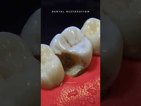
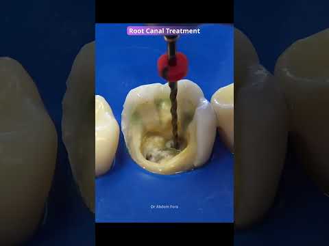
A cause of endodontic failure: failure to locate of the Middle Mesial Canal MMC. During root canal treatment—particularly on upper and lower first molars—an additional canal is often present; in the case of lower molars, this canal is located in one of the roots and is known as the "middle-mesial canal". This canal must be located and disinfected before being obturated to ensure the success of the endodontic treatment. Join me as we look at a case study, covering everything from caries removal to the complete restoration of the tooth. Importance of Middle Mesial Canal 🦷 -Prevalence: Reported in roughly 1% to 28% of mandibular first and second molars depending on the detection methods and population studied. -Detection: Often hidden beneath a small wall of dentin. Dentists use advanced tools like Cone-Beam Computed Tomography (CBCT) and surgical microscopes to spot them. -Risk: Missing this hidden canal during root canal therapy can leave bacteria behind and lead to treatment failure. Captions: 0:00 Intro 0:49 Caries Removal 2:19 Absolute isolation with rubber dam 2:25 Deroofing the pulp chamber 2:51 Endodontic canal patency 3:05 Shaping and cleaning of the mesiobuccal canal MB 4:04 Shaping and cleaning of the mesiolingual canal ML 4:26 Irrigation with sodium hypochlorite and EDTA 5:39 Location of the Middle Mesial Canal MMC 5:54 Shaping and cleaning of the Middle Mesial Canal MMC 6:56 Shaping and cleaning of the Distal canal D 7:47 Drying of canals with paper points 8:05 Root canal obturation with gutta-percha 9:46 Fiber post preparation 10:14 Tofflemire Matrix 10:51 Fiber Post cementation 11:32 Composite resin core buildup 12:28 Overlay preparation 13:36 Analog impression taking 14:04 Overlay 14:32 Overlay conditioning 15:19 Tooth conditioning 15:36 Overlay cementation 15:54 UV light activation 16:03 Polishing 16: 35 Before and after Leave a comment, like, and share this video!